Cardiac a rrhythmias can also be classified according to their location, ie, depending on the area of the heart where the arrhythmia is initiated. In this case we talk of:
Supraventricular arrhythmias, which originate in the atria or any cardiac structure
Ventricular arrhythmias, when they are generated in the ventricles. These arrhythmias are the most dangerous as they directly affect the ability of the heart to pump blood to the rest of the body. In fact, ventricular tachycardia and ventricular fibrillation are the main arrhythmias leading to sudden cardiac death.
1. Bradycardias
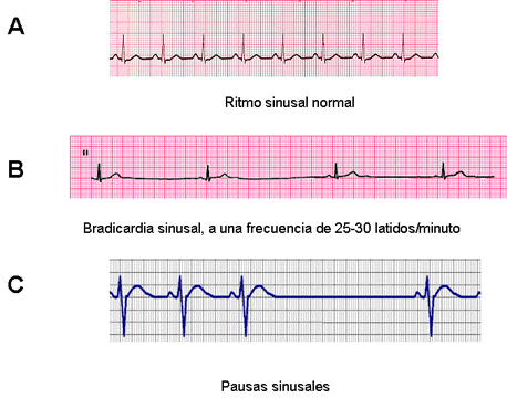
Bradycardias are those situations in which the heart beats at a frequency slower than normal, i.e., less than 60 beats per minute. Bradycardia can occur by decreasing the frequency at which pulses are generated in the sino- atrial node (sinus bradycardia) or because the impulses which are generated at this level can not stimulate the whole heart because they are blocked at some point of the conducting system (it is indicated in colours in the figure) that conduct electrical impulses through the heart. In this case we talk about intracardiac blocks. The points at which the impulses are most often blocked are the atrioventricular node (which is the only pathway though which impulses pass from the atria to the ventricles) and the bundle of His. In the Figure we indicate with thick blue the atrioventricular node and the His bundle.
Sinus bradycardia Healthy and elderly people sometimes present cardiac frequencies below 50 bpm. Healthy trained athletes and young adults at rest or at night can present low heart rates (even of less than 30 bpm) without symptoms or an increased cardiovascular risk. Various diseases (myocardial infarction, hypothyroidism, intracranial hypertension) and certain drugs (amiodarone, morphine, beta-blockers, verapamil, diltiazem, digoxin) can produce sinus bradycardia. It also appears when there is a predominance of vagal tone (vasovagal syncope, vomiting, abdominal surgery). Sometimes sinus bradycardia is due to the aging or to a disease leading to a degeneration of the sino-atrial node.
Sinus b radycardia rarely produce symptoms if the frequency is no lower than 50 bpm. When the frequency is lower than 50 bpm symptoms can appear because the heart does not pump enough blood towards the various tissues of the body. This may cause dizziness, weakness, instability, fatigue, tiredness, low blood pressure, fainting (feeling of impending fainting) or syncope (fainting or loss of consciousness).
Bradycardia or asymptomatic intracardiac blocks do not require treatment. If the bradycardia is symptomatic, treatment should be aimed at controlling the triggering causes. If a drug is the cause, the suppression of treatment may be sufficient. When bradycardia or intracardiac blocks produce symptoms (e.g. syncope), a permanent pacemaker should be implanted.
Sinus pauses. In this case the heart is not activated, either because pulses are not generated in the sino- atrial node or because the pulses generated at this level are blocked at some point in the atrium. The pauses usually last for several seconds and can cause recurrent syncope. An implantable pacemaker is recommended in patients with pauses of 3 or more seconds.
Sick sinus syndrome (sinus node dysfunction).This is a cardiac arrhythmia presumably caused by a malfunction of the sino-atrial node, the heart's pacemaker. This arrhythmia is related to disorders that cause scarring, degeneration, or damage of the cells forming the sinus -atrial node due to cardiac diseases leading to fibrosis, myocarditis o amiloidosis. Thus, it is more frequent in elderly people and often is intermittent. Patients present bradycardia which dizziness, palpitations, fatigue, chest pain, unsteadiness, loss of concentration, irritability or fainting (syncope) due to cardiac arrest (asystole). In these patients is necessary to implant an artificial cardiac pacemaker.
Bradycardia-tachycardia syndrome, is a variant of sick sinus syndrome in which slow and fast heart artes alternate.
Atrio-ventricular (AV) conduction block In this case, the pulses that have invaded the atrium are conducted with delay or they are not conducted through the atrio-ventricular node to the ventricles. The blockade of impulse conduction may occur in the AV node or in the His bundle and in its branches. AV block can occur in trained young athletes during sleep. It can also be p roduced by some drugs (digoxin, beta -blockers, verapamil, diltiazem, amiodarone), infections (viral infections, rheumatic fever) or various heart diseases (e.e., inferior myocardial infarction). Arterial hypertension and mitral or aortic valve stenosis are associated with increased fibrosis and calcification of the heart, which can lead to degeneration of the AV node or the His bundles and facilitate the development of block at both levels.
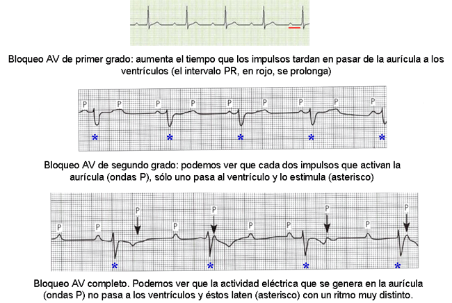
First-degree atrioventricular (AV) block. This means that the impulse conduction from the atria to the ventricles through the AV node is delayed. This is evident in the electrocardiogram as a PR interval greater than 200 ms in length. The blockade can be due to an increase in vagal tone (athletes), myocarditis, acute inferior myocardial infarction and drugs that slow the conduction through the AV node (calcium channel blockers,beta-blockers,cardiac glycosides). Patients with an isolated first degree AV block usually are asymptomatic, although symptoms may occur especially during exercise. Thus, it has a benign prognosis. The management includes withholding any offending drug. Clinicians should be very cautious when introducing any drug that may slow AV conduction and regular monitoring of the ECG is indicated.
Second-degree atrioventricular (AV) block is a disorder characterized by delay, or interruption of atrial impulse conduction to the ventricles through the atrioventricular node. In the ECG we can see that some P waves (an indication of atrial electrical activity) are not followed by a QRS complex which represents the ventricular activation. The AV block can be permanent or transient.
a) Mobitz I second-degree AV block, characterized by a progressive prolongation of the PR interval. Ultimately, the atrial impulse fails to conduct (the QRS complex is absent), and there is no ventricular contraction. It can be due to an enhanced activation of the parasympathetic nervous system, such as in well-trained athletes (it appears in 2-10% of long distance runners) or during sleep. The conduction delay generally occurs in the AV node (70%) or infranodally (30%). Most patients are asymptomatic (particularly well-trained athletes), although light-headedness, dizziness, or syncope can appear in patients with structural heart disease.
Vagally mediated AV block has a good prognosis from a mortality standpoint but may lead to dizziness and syncope. When the AV block is localized to the AV node and there is no organic heart disease there is no increased risk of morbidity or death, while block appearing during an acute myocardial infarction can increase mortality. The risk of progression to complete heart block increases when the level of block is in the His-Purkinje conduction system (infranodal).
Asymptomatic patients do not require any specific therapy. Symptomatic patients should be treated with atropine and transcutaneous pacing (particularly those with an infranodal blockade). Atropine improves conduction through the AVN by reducing vagal tone. This explains why it is unlikely that patients with an infranodal block can benefit from atropine. Additionally, atropine should be administered with caution in patients with suspected myocardial ischemia, as ventricular dysrhythmias can occur. Patients with suspected myocardial ischemia should be treated as appropriate. In drug-induced AV block a decrease in the dose or the discontinuation of the medications (digoxin, diltiazem, verapamil, b -blockers) may restore normal AV conduction.
b) Mobitz II second-degree AV block, characterized by an unexpected non-conducted atrial impulse, without prior measurable lengthening of the conduction time (i.e., here the PR interval is constant). Here, the conduction delay generally appears infranodally. Mobitz type II blocks carry a risk of progressing to complete heart block, and thus are associated with an increased risk of mortality.
Patients are more likely to experience light-headedness, dizziness, or syncope; chest pain can be present if the heart block is related to myocarditis or ischemia. Bradycardia and signs of hypoperfusion, including hypotension may be present.
A transvenous pacemaker is indicated, even in asymptomatic patients. An anti-ischemic regimen should be instituted if ischemia is suspected.
Third-degree (complete) AV block. Under these circumstances the impulses generated in the SA node in the atrium does not propagate to the ventricles. Thus, the ventricles are activated by an accessory ventricular pacemaker. Some patients present minimal symptoms (dizziness, weakness, or malaise); on the contrary, other patients present bradycardia,hypotension, hert failure, cardiovascular collapse, or death.
The most common cause of complete AV block is coronary ischemia. An inferior wall myocardial infarction may cause a third-degree heart block that usually resolves within 2 weeks. An anterior wall myocardial infarction may damage the distal conduction system of the heart, causing third-degree heart block associated to a permanent damage to the conduction system.
Third-degree AV block can be treated by use of an artificial pacemaker. It is encouraged the treatment of other diseases associated with AV block, such as myocardial ischemia. Atropine is not effective for treating 3rd degree blocks.
Intraventricular conduction abnormalities
Under ceratin conditions impulse conduction can be slowed or stop in the ventricles. Quite often the block is located on the right or the left branch of the His bundle and thus, we say that the patient has a bundle branch block. This happens frequently in the elderly and in patients with heart diseases (myocardial infarction, heart failure, long-standing hypertension, cardiomyopathies or a disease of the heart valves). The right bundle branch block can occur occasionally even in a healthy heart, while the left bundle branch block usually occurs in patients with an increased risk of cardiovascular diseases.
The cardiac electrical impulse must be propagated at the same speed through the right and left branches of the His bundle, so that the right and the left ventricles contract simultaneously. However, if there is a blockage in one of these branches, the electrical impulse reaches the ventricle through an alternative pathway and although the rate and rhythm of the heart are not affected, the impulse spreads more slowly than usual, so the ventricles are not able to contact simultaneously, but in one of them the contractile process is going to be a little more slowly than in the other.
The bundle branch block can be chronic or intermittent and occasionally the blockade does not produce symptoms. If so, the patient does not require any treatment. Conversely, the bundle branch block can produce dizziness, fainting, feeling of fainting (syncope) or near syncope. In symptomatic patients, particularly if the bundle branch block is associated with cardiovascular diseases, is necessary to implant a pacemaker to facilitate that both ventricles contract simultaneously. In patients with bundle branch block and dilated cardiomyopathy, a new type of cardiac stimulation called"cardiac resynchronization therapy (CRT)"that allows to coordinate the beating of both ventricles, avoiding cardiac dyssynchrony, by simultaneously pacing both the left and right ventricles.
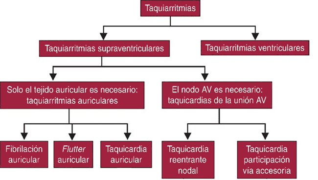
Tachycardia (or tachyarrhythmia) means that heart rate is rapid, above 100 bpm. The arrhythmia often appears under circumstances characterized by an increase in sympathetic tone (fear, stress, exercise), in the presence of fever, heart failure or hyperthyroidism or after administration of stimulants (coffee, tea, snuff) and some drugs. Tachycardias occur with rapid irregular heartbeat in the chest, chest discomfort, weakness, shortness of breath, sweating, and dizziness.
Supraventricular Tachyarrhythmias
They are generated in the atria or the atrioventricular node. The patients p resent palpitations, anxiety, dizziness, sweating, breathlessness, anxiety, low blood pressure, presyncope (characterized by lightheadedness, muscular weakness, and feeling faint) and sometimes syncope. These symptoms are due to the fact that at rapid frequencies (above 140 bpm) the diastolic interval is markedly shortened and the heart is not adequately refilled with blood and, therefore, the amount of blood pumped to the tissues decreases. Tachyarrhythmias can also cause and/or aggravate signs of coronary ischemia, can produce chest tightness and angina (chest pain caused by lower blood supply and oxygen to the heart).
Atrial tachycardia. This is an arrhythmia that originates in the atria, sometimes in one place (focal tachycardia) or in several places simultaneously (multifocal tachycardia). The focal tachycardia responds very well to radiofrequency ablation.
Atrial fibrillation. Atrial fibrillation (AF) is a heart condition that causes an irregular, uncoordinated and abnormally fast heart rate (> 350 bpm). It is the most common sustained arrhythmia, occurring in 1-2% of the general population, and accounts for approximately one-third of hospitalizations for cardiac rhythm disturbances and is related to considerable morbidity, mortality and economic burden. It is the most common cardiac arrhythmia, occurring in 1-2% of the general population. The risk of AF increases with age, from 0.5% at 40-50 years to 5-15% at 80 years. AF represents a significant burden to patients, ranging from the impact of debilitating symptoms on daily life to the increased risk of stroke and/or death. In fact, AF is associated with a nearly doubled risk of death and an almost 5-fold increase in the risk of stroke. Learn more
Wolff-Parkinson-White syndrome. In some patients there are abnormal accessory electrical conduction pathway s between the atria and the ventricles. Under these conditions, atrial impulses can travel from the atria to the ventricles: through the atrioventricular node or through these abnormal pathways. The risk is that if these patients present a rapid atrial rate (eg atrial fibrillation) the accessory bundle may conduct all the electrical impulses from the atria to the ventricles, producing a n excessive increase in the ventricular rate, leading to ventricular tachycardia or ventricular fibrillation, hypotension, cardiorespiratory arrest and sudden death of the patient.
Patients with rapid ventricular arrhythmias may require a synchronized electrical cardioversion. If they are relatively stable, antiarrhythmic drugs (amiodarone, flecainide, propafenone) may be used. The definitive treatment of WPW is a destruction of the abnormal electrical pathway by radiofrequency catheter ablation.
These tachyarrhythmias are originated and maintained in the ventricles of the heart, distal to the bundle of His.
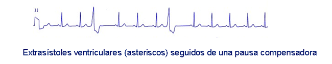
Ventricular extrasystoles. They are premature ventricular contractions interrupting the normal heart beats, which are originated from a source which is located in the ventricles, distal to the bifurcation of His bundle. They occur in the elderly and in patients with an excessive consumption of coffee, tobacco abuse, emotions, stress; in patients with coronary artery disease (after myocardial infarction), cardiomyopathes, hypopotasemia, infections or hyperthyroidism.
Symptoms. Include palpitations, chest discomfort, feeling of heart stopping, followed by a stronger beat, faintness, syncope. Treatment. Ventricular extrasystoles should be treated only if they are symptomatic. Ventricular extrasystoles that appear on a healthy heart require the removal of potential triggers (nicotine, caffeine, diuretics, cocaine, sympathomimetics). I f they occur in patients with heart disease it is necessary to correct electrolyte and acid-base disorders, myocardial ischemia and other possible factors implicated in their appearance. Antiarrhythmic drugs can be prescribed in symptomatic patients. Beta blockers and amiodarone are the first choice drugs.
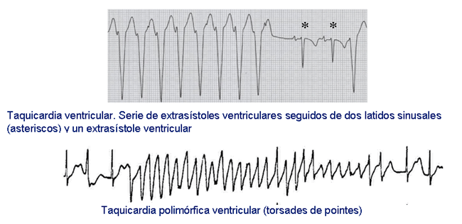
Ventricular Tachycardia (VT). It is defined as a series of three or more ventricular complexes occurring at a frequency greater than 120 bpm (up to 250 beats/min). The VT a ppears in most of the cases in patients with structural heart disease. In fact, they occur in 10-40% of patients with myocardial infarction. They also appear in patients with heart failure, cardiomyopathy (diseases specific of heart muscle), valvulopathy (diseases of the heart valves) or cardiac surgery or may be caused by drugs.
Symptoms. Symptoms depend on the ventricular rate, duration of arrhythmia and the presence and gravity of cardiovascular disease modifying the function of the ventricles pump. If the VT se lf-terminates within 30 seconds, it is considered a non-sustained VT. If the arrhythmia lasts more than 30 seconds, it is known as a sustained VT.
When the ventricles beat in a very fast rates (140-220 bpm) for more than 30 seconds (sustained VT) is not enough time to fill with blood the ventricles during diastole (period when the heart relaxes after a contraction) and, therefore, decreases the volume of blood they pump to the arteries. A sudden increase of ventricular rate during VT leads to the appearance of a sensation of feeling the heart beat (palpitations), dizziness, vertigo, signs of heart failure (shortness of breath or dyspnea), chest pain or chest tightness and unconsciousness. Blood pressure is usually low and pulse absent. Sometimes non-sustained VT is so brief that hardly produces symptoms. Conversely, in some patients the VT persists over time and degenerates in ventricular fibrillation, and cardiac arrest associated with the sudden death of the patient. Treatment. Asymptomatic patients should be advised to suppress the consumption of alcohol, caffeine and other stimulant substances or drugs that can induce or increase the frequency of ventricular premature beats. If this is not enough, an antiarrhythmic drug should be prescribed. Drug selection should be based on drug safety and on the structural heart disease of the patient. After an acute myocardial infarction may be administered lidocaine, procainamide, beta-blockers or amiodarone. When the patient does not respond to treatment or if the TV produces hypotension, angina, heart failure or signs of cerebral hypoperfusion, electrical cardioversion should be performed immediately.
Electrical cardioversion (also known as direct-current or DC cardioversion) is a procedure whereby a synchronized electrical current (shock) is delivered through the chest wall to the heart through special electrodes or paddles that are applied to the skin of the chest and the back. The purpose of the cardioversion is to interrupt the abnormal electrical rhythms in the heart and to restore a normal heartbeat. The delivered shock causes all the heart cells to contract simultaneously, thereby interrupting and terminating the abnormal electrical rhythm without damaging the heart. The heart's electrical system then restores a normal heartbeat.
In patients where it is suspected that VT can degenerate into ventricular fibrillation and cause a sudden cardiac death an automatic implantable defibrillator should be implanted. This is a small battery -powered electrical impulse generator that when detects that a ventricular tachycardia occurs discharge an electric shock to the heart usually returns to its normal rhythm.
To prevent the occurrence of new episodes of VT it is necessary to treat aggressively the coronary artery disease and the heart failure, as both processes facilitate the appearance of the TV. In most patients the chronic antiarrhythmic drug therapy of VT includes amiodarone and beta -blockers.
Fibrilación ventricular. This is the most serious (malignant) cardiac rhythm disturbance characterized by multiple, rapid (> 250 bpm) and irregular electrical activity of the ventricles. As a consequence, the ventricles do not pump any or pump little blood to your vital organs, leading to loss of consciousness (syncope), seizures, cardiac arrest and sudden cardiac death. Cardiac arrest is diagnosed when a person suddenly collapses, turns deathly pale, has very dilated pupils, and has no detectable pulse, heartbeat, or blood pressure. Ventricular fibrillation is typically the first expression of coronary artery disease (CAD) and is responsible for approximately 50% of deaths in patients with CAD.
The following symptoms may develop before any major cardiac event: chest pain and other angina equivalents, dyspnea, fatigue, palpitations, s yncope and immediately preceding acute cardiac arrest, an increase in heart rate, presence of premature ventricular contractions or a period of VT.
VF is fatal unless treated immediately. Cardiopulmonary resuscitation must be started as soon as possible followed by defibrillation, which gives an electrical shock to the heart. People who are successfully resuscitated from ventricular fibrillation and survive are at high risk of another episode. Thus, if ventricular fibrillation is caused by a reversible disorder, this should be treated. Antiarrhythmic drugs may then be given to help maintain the normal heart rhythm.
Sudden cardiac death. Sudden cardiac death (SCD) is one that occurs suddenly and unexpectedly in the first hour after the onset of symptoms in an individual who may or may not have a history of heart problems. It is the most common cause of death in the Western countries and, in the EEUU, it accounts for 250,000-400,000 cases annually, i.e., more than breast cancer, lung cancer and HIV/AIDS combined. Thus, around 12.5 % of deaths that occur naturally are sudden. Of the total cases of SCD, almost 80% occur in patients with ischemic heart disease or heart f ailure. Another 15 % occurs in patients with structural heart disease, eg cardiomyopathie s. Finally, in less than 5% of patients, SCD occurs in healthy young individuals with a normal heart with mutations in the genes encoding cardiac ion channels, i.e., these patients present genetically-based or inherited cardiac arrhythmias.
The most common cause of SCD is a ventricular tachycardia that degenerates into ventricular fibrillation, loss of consciousness and cardiac arrest. Circulatory arrest is accompanied by cessation of breathing movements (sometimes may persist briefly gasping respiratory movements), generalized convulsions and fixed and dilated pupils, and sometimes, a marked cyanosis (bluish discoloration of the skin and mucous membranes).
Most patients who suffer SCD and do not receive medical attention will die within minutes. T herefore, the best way to prevent SCD is to identify those patients at high risk of developing an episode of SCD. Since the majority of high-risk patients are those with a previous myocardial infarction, it is necessary to perform a strict medical control in these patients in order to improve the supply of oxygen and nutrients to the heart.
The treatment of choice is the cardiac electrical defibrillation while quickly begin the maneuvers of cardiopulmonary resuscitation. Since the patient can recover by cardiopulmonary resuscitation, the MSC may be recurrent.